
Neurodevelopment //
Neurodevelopment Research at MRN
A neurodevelopmental disorder is one that impairs a child’s ability to develop along a typical trajectory. While the symptoms and causes of these disorders can vary widely, targeted interventions generally lead to better outcomes for these children. Since the brain is most malleable in children less than five years of age, the earlier interventions begin, the more likely these children will follow a closer to normal developmental path. This will hopefully lead to improved quality of life for these children and their families by increasing the child’s independence as well as reducing long-term medical costs. One of the barriers to early identification is that many disorders are diagnosed behaviorally, which is limiting due to the natural variability in behavior among healthy children. Behavioral data often fail to indicate which interventions will be most appropriate for a given child, so understanding the atypical brain responses may help to develop and guide the use of a specific treatment.
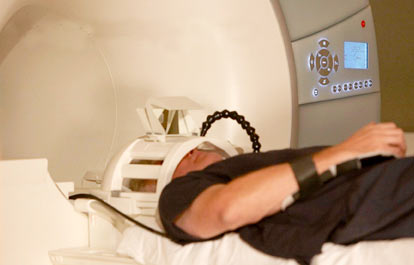
Using noninvasive imaging, MRN investigators are studying brain development from birth through childhood with the goal of using brain structure and function to identify markers of disorders for the purpose of guiding therapies. Beginning with specific disorders, progress has been made on several fronts, including mapping epileptic activity in young children, and identifying structural and functional differences in children born prematurely. Using the world’s first pediatric MEG system (the babySQUID®), Dr. Julia Stephen and her team have identified a measure of atypical brain connectivity in children as young as 20 months who have been diagnosed with an autism spectrum disorder (ASD). Identification of specific markers in children already diagnosed with an ASD may help identify markers of atypical brain development in young children who may be at risk for developing this disorder. Dr. Stephen is also focusing on identifying markers in children aged three to five years with fetal alcohol spectrum disorders (FASD). Due to the stigma associated with drinking alcohol during pregnancy, many children are not diagnosed with FASD until as late as five years of age. Like ASD, early intervention is key to improving the quality of life of children with an FASD, who are known to be at higher risk for developing other mental disorders. Please visit Dr. Stephen's Child Study website.
With funding from the Delle Foundation, Drs. Arvind Caprihan and John Phillips are collaborating on a large scale project to investigate how MRI can be used to better understand normal brain development in children from birth to five years of age. In addition to performing a normative longitudinal study on children in the first year of life, Drs. Caprihan and Phillips are collaborating with University of New Mexico researchers on a new and important NIH study to better understand the structural and functional changes associated with providing erythropoietin to premature children in neonatal intensive care.
The neurodevelopment team also includes senior investigator Dr. Jeffrey Lewine. His autism research focuses on identifying markers that will help identify optimal intervention techniques for individual children. He is currently conducting a tinnitus study examining the safety and efficacy of a new procedure for reducing tinnitus - Tinnitus Re-Organization Training (TROT). Through these combined efforts, MRN neurodevelopment researchers are working toward improving the identification of developmental disorders and optimizing interventions based on known anatomical and/or functional deficits.
Current Research //
- Comprehensive Evaluation in the Relationships of Early Brain Response Overtime (CEREBRO) Project >
- Brain Imaging and Developmental Follow-up of Infants Treated with Erythropoietin (BRITE) Project >
- Kids, Imaging and Developmental Outcome (KIDO) Project >
- Identifying Markers of Atypical Brain Development Due to Prenatal Alcohol Exposure >
- Executive Function and Early Child Development in Healthy and Preterm Infants >
- Biomarkers for White Matter Injury in Vascular Dementia >
- Premature Infant Development >
- Biomarkers for White Matter Injury in Mixed and Vascular Cognitive Impairment >
Comprehensive Evaluation in the Relationships of Early Brain Response Overtime (CEREBRO) Project
This is a longitudinal study of normal brain development from 4 months of age through the preschool years. Children are followed with serial assessments including developmental tests, MRI scans and genetics. Our developmental assessments include a focus on early childhood self-regulation and executive function development. By following this cohort of children over time we hope to characterize individual differences in normal brain development, which offers an opportunity to study brain-behavioral relationships in both healthy children as well as those at risk of developmental disorders.
Brain Imaging and Developmental Follow-up of Infants Treated with Erythropoietin (BRITE) Project
This is the first study that uses neuroimaging to study the neurologic consequences of erythropoietin treatment for very premature infants. We follow three groups of children: 1) children born prematurely who were treated in the newborn intensive care unit with erythropoietin, 2) similar premature children who were not treated with erythropoietin, and 3) healthy term-born children. These children are evaluated at approximately 3 years of age and again at approximately 6 years of age with MRI scans and developmental assessments. Our goal is to determine whether early erythropoietin therapy improves developmental outcome, and secondly to characterize the neurologic mechanisms of that improvement.
Kids, Imaging and Developmental Outcome (KIDO) Project
Some, but not all, children born after toxic exposures during pregnancy grow to have developmental and behavioral challenges. Treatment is more effective if begun early, however identifying children at greatest risk is difficult until clinical problems have begun to occur. We are beginning to study children born after in utero toxic exposures with neuroimaging and developmental assessments beginning in the first months of life in an effort to better understand the neurologic mechanisms resulting in cognitive and behavioral delay. We hope this work will lead to improved approach to early diagnosis and treatment of these children.
Identifying Markers of Atypical Brain Development Due to Prenatal Alcohol Exposure
Prenatal alcohol exposure is the leading cause of developmental delays in children. However, due to the stigma associated with drinking during pregnancy, it is often difficult to identify the children at risk of developmental delays due to prenatal alcohol exposure. Identification of prenatal alcohol exposure at a young age provides the opportunity for early intervention to optimize long-term outcomes. Left untreated, these children are at higher risk of developing addictions and being incarcerated than controls. Based on their neurobehavioral profile, identification of prenatal alcohol exposure in adolescence would suggest a different course of treatment for at-risk youth and could prevent long-term incarceration. This NIH funded pilot study uses MEG, EEG and MRI to identify a unique pattern of brain dysfunction in children prenatally exposed to alcohol.
Executive Function and Early Child Development in Healthy and Preterm Infants
Neuroimaging imaging measures to serve as predictive biomarkers of executive function development in healthy and preterm infants are being developed. This is based on integrating changes in cortical development, white matter tracts, cerebral blood flow, and proton spectroscopy with developmental assessments.
Biomarkers for White Matter Injury in Vascular Dementia
White matter biomarkers based on blood-brain disruption and water diffusion in the white matter are being developed to characterize progression of white matter hyperintensities and to identify patients with Alzheimer’s and subcortical ischemic vascular disease who can develop cognitive impairment.
Premature Infant Development
Enormous and rapid changes in the brain occur during the neonatal period and first years of our lives, which makes the brain particularly vulnerable to physiologic stresses such as premature birth.
In the project BRain imaging and developmental follow-up of Infants Treated with Erythropoietin (BRITE), led by Drs. Robin Ohls and John Phillips, we are investigating the impact of erythropoietin (Epo) treatment on brain development in young children who were born prematurely (with very low birth weight or VLBW). Our preliminary results on altered neurochemistry in prematurely born children is reported in JP Phillips, et al., Anterior Cingulate and Frontal Lobe White Matter Spectroscopy in Early Childhood of Former Very Low Birth Weight Premature Infants, Pediatr Res. 69(3):224-9 (2011). We observed that both NAA and total creatine are on average low in VLBW children relative to normal birth weight children, suggesting reduced neuronal function or density along with a reduced energy metabolism in VLBW children. These findings were consistent with a lower performance on cognitive tests demonstrated by the VLBW group. Our hypothesis is that follow-up studies on these children 2 years later and after treatment with Epo will reveal a normalization of these biochemical and cognitive markers towards the levels in the healthy control group.
Biomarkers for White Matter Injury in Mixed and Vascular Cognitive Impairment
Another fate that awaits most of us, as we enter the latter decades of our lives, is cognitive decline. Whether this is a normal loss of memory or cognitive “processing speed” that we learn to manage and live with or a more serious loss of cognitive function, as in Alzheimer’s disease or any of the many forms of vascular dementia, depends on factors that are not well understood at present.
The project Biomarkers for White Matter Injury in Mixed and Vascular Cognitive Impairment, led by Dr. Gary Rosenberg, is aimed at combining several clinical and neuroimaging measures to arrive at a better differential diagnosis of vascular dementia, to help guide the development of treatments, as well as to discover more about the underlying pathologies of the different forms of the disease. Early results from this project have been reported in Taheri et al., Blood-Brain Barrier Permeability Abnormalities in Vascular Cognitive Impairment, Stroke, 42(8):2158-63 (2011). A preliminary report of our neurochemical findings (Gasparovic et al., submitted, 2012) demonstrates that NAA and total creatine are more strongly related to cognitive function in subjects with vascular dementia than is ischemic lesion volume.

